


Glomus tumor: why can such a small lesion cause so much pain?

A lesion measuring just a few millimeters can cause surprisingly intense pain. This is one of the signs of a glomus tumor, a rare and generally benign growth. While most commonly found under the fingernail, it can also appear in other parts of the body, including the knee.
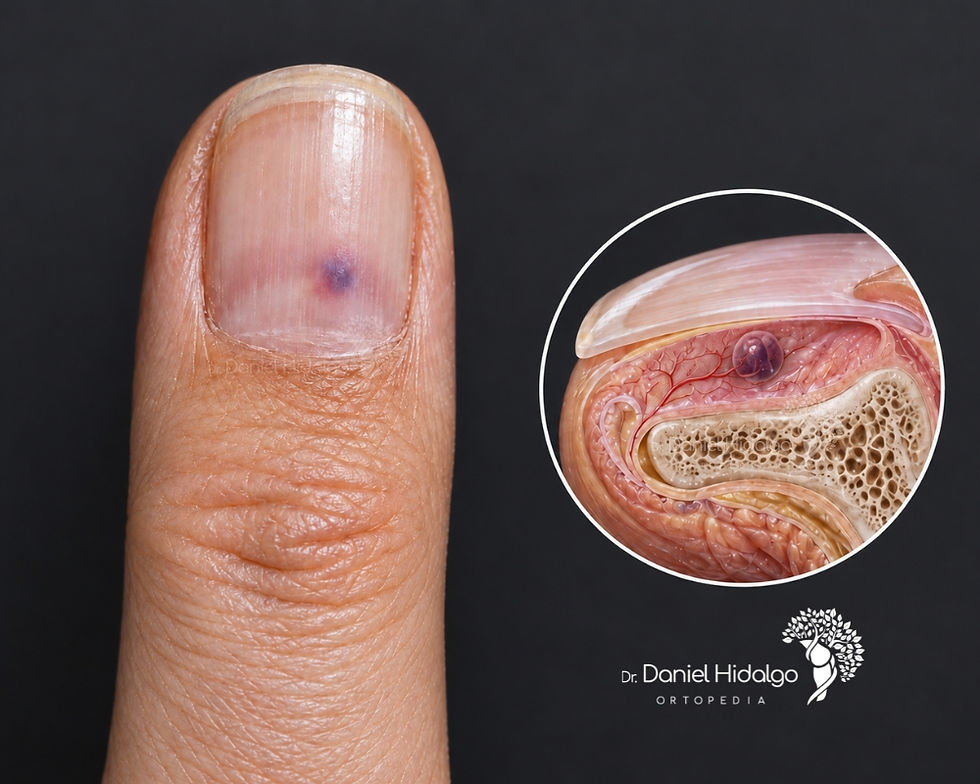
What is the glomus body?
The glomus body is a small skin structure that helps regulate temperature by controlling blood flow. It is especially abundant in the fingertips. A glomus tumor develops from cells similar to those of this structure. It is not a tumor of a nerve receptor, although it can cause intense pain.
What are the symptoms?
The most characteristic signs are localized pain, intense sensitivity to pressure at a specific point and worsening with cold. When it is under the nail, the tumor may produce a small bluish or reddish spot, but it is not always visible. Not all patients present with all three symptoms, especially in locations outside the fingers.
Can it appear on the knee or in other areas?
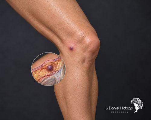
Yes. In this situation, it is called an extradigital glomus tumor. There are published reports of cases occurring in the front of the knee and in other structures near the joint. Because this location is uncommon, the cause of the pain may go unnoticed for a long time. A small area that is very painful to the touch—sometimes accompanied by a nodule—warrants evaluation.
How is the diagnosis made and what is the treatment?
The investigation begins with the patient's symptom history and an examination of the painful area. Ultrasound and magnetic resonance imaging (MRI) can help locate the lesion and plan its removal, particularly in unusual locations. Confirmation is typically achieved through pathological examination (biopsy). When symptoms are present, the standard treatment is complete surgical removal, which can provide pain relief. The decision depends on the assessment of each individual case.
About the author
Dr. Daniel Hidalgo is an orthopedic surgeon specializing in knee surgery, providing diagnosis as well as conservative and surgical treatments for injuries.
If you are experiencing knee pain, instability, or limitations in your activities, please contact us for a specialized evaluation. This will allow you to clarify your diagnosis and determine the best treatment plan.
Learn more
The images in this post are labeled as illustrative, were created with the help of artificial intelligence, and do not represent the diagnosis of a specific patient.


Comments