
Patellar Instability
The patella is the circumferential bone located at the front of the knee. It connects the strong quadriceps muscle, which is located at the front of the thigh, to the patellar tendon.
In the posterior aspect of the patella, there is a layer of cartilage that sits in close contact to the cartilage surface at the front of the femur, in a area called the trochlea. The joint between the patella and the trochlea is called the patellofemoral or femoropatellar joint.
In a normal situation, the patella should remain in congruence with the trochlea during all knee movements. However, on certain occasions, the patella can move outwards the knee, no longer having contact with the trochlea. This phenomenon characterizes patellar dislocation.

When It Happens
Patellar dislocation can occur after a torsional trauma to the knee, in which the foot becomes trapped and rotates outwards while the knee is being leveraged. The dislocation causes intense joint pain ad effusion and the patient usually needs to be taken to the hospital for a medical evaluation. Deformity, swelling and difficulty in moving the knee may occur.
In some patients, the patella can dislocate without a significant twisting of the knee and this can occur repeatedly. In these cases, the first dislocation usually occurs in childhood and the patients can be diagnosed with patellar instability.
Patellar instability or patellofemoral instability is more common among young girls, aged 10 to 17 years. Patients with patellar instability usually have anatomical alterations that favor the dislocation. The main anatomical factors related to instability are:
-
Trochlea dysplasia;
-
Valgus alignment of the extensor mechanism;
-
Increased patellar tilt;
-
High patella.
There are also some secondary factors related to patellar instability. They are:
-
Excessive femoral anteversion;
-
Excessive external rotation of the tibia;
-
Knee bow;
-
Knee valgus.
Diagnosis
Patellar dislocation is usually quite painful, requiring quick medical intervention to put the patella back in its usual place.
The patient usually tells the doctor how the dislocation occurred. Patients with recurrent dislocation usually find it easier to report.
The diagnosis of a dislocated patella can be made clinically, since the deformity can be easily observed by the doctor. However, when the patient comes to the doctor with the patella in place, when the patella returns to its usual place involuntarily, the diagnosis may not be so easy.
Patients with patellar instability should be examined carefully, since there are several possible changes in the exam that can indicate the causes of the patellar dislocation.
In addition, some exams should be requested by the orthopedic surgeon so that the anatomical changes related to the patellar instability can be observed.
The exams that provide the most information are:
-
Knee X-rays;
-
Computed tomography;
-
Nuclear magnetic resonance.
Treatment
After the first episode of patellar dislocation, still in the acute phase of the problem, treatment must be immediate so that the patella can be put back in its usual place. Often, the patient comes to the doctor with the patella already in place.
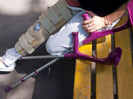
After certifying that the patella is in the correct position and that no complications have occurred, such as fracture, intra-articular free fragment, among others; non-surgical treatment is indicated, based on anti-inflammatory and analgesic medications, ice on the front of the knee and immobilization with a plaster cast or removable device. The period of immobilization varies from two to six weeks. In the acute phase, it may still be necessary to puncture the knee for pain relief if the swelling or joint effusion is very significant.
After immobilization, the patient should be referred for rehabilitation with physiotherapy.
The treatment of patients with patellar instability, with recurrent episodes of dislocation, is different. It is based on motor physiotherapy to rebalance the muscles involved and on the surgical correction of the anatomical factors related to patellar instability.
There are several surgeries described for the treatment of patellar instability. However, the most frequently performed procedures are:
-
Reconstruction of the medial patellofemoral ligament (MPFL);
-
Chondroplasty or osteochondroplasty (arthroscopic or conventional);
-
Osteotomy of the tibial tuberosity;
-
Release of the lateral retinaculum of the patella (arthroscopic or conventional).
-
After surgical treatment, it is important that the patient immediately begins rehabilitation with motor physiotherapy.